
")
RFLSI III

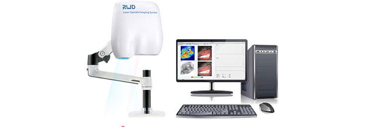
RFLSI Ⅲ is based on the LSCI (Laser Speckle Contrast Analysis Imaging ) technology design, non-invasive with non-contact, high time resolution, high spatial resolution, and full-field rapid imaging. It provides real-time dynamic blood flow monitoring and recording for research, perfusion evaluation, such as MCAO, cerebral blood flow. compared with lase doppler imaging, it can realize full field video imaging function.
Applications:
- Cerebral blood flow measurement, CBF, blood vascular
- MCAO mouse/rat model or photostromboic stroke models
- Cortical spreading depression (CSD)
- Traumatic brain injury (TBI)
- HLI/CLI, the middle ear of rodent, burn assessment, etc.
- Animal monitoring during brain surgery.
Features:
- Analyze vascular perfusion volume, tube diameter, tube angle, and other data online and offline to meet hemodynamic research. HD Blood flow meter.
- Multiple data acquisition methods including continuous acquisition, specified interval acquisition, specified frame number acquisition, etc.
- Indicating laser plus autofocus function makes it easy to use.
- High-definition camera (2048 × 2048 pixel) helps you see the end of blood vessels and open the world of blood vessel microcirculation images.
- The high-speed camera (up to 120 FPS) allows us to record changes every second and record more details of blood vessel changes after intervention.
- Package-type supporting solutions and timely after-sales service will help you to be one step ahead in your scientific research.
- Easy to use, USB 3.0 connection, flexible and simple setup helps get you up and running quickly.
Laser Speckle Imaging System (RFLSI Ⅲ) is a blood perfusion imager based on Laser Speckle Contrast Imaging (LSCI) technology. LSCI provides a better means to study the microcirculation in a way that was not possible in the past. It allows visualization of tissue blood perfusion and imaging with high time and spatial resolution. There is no influence on the perfusion, as no contact to the tissue is needed, nor dyes or tracer elements.
What is Speckle Contrast Analysis?
Speckle flow techniques are based on the changes over time of the dynamic speckle pattern generated by motion in the sample. In these techniques, this changing speckle pattern is recorded with a camera that has an integration time in the order of the speckle decorrelation time (i.e., in the millisecond range). Due to the long integration time compared to the typical decorrelation time of the speckle pattern, the speckle pattern will be blurred in the recorded image. The level of blurring is quantified by the speckle contrast1.
1.Draijer, M., Hondebrink, E., van Leeuwen, T. & Steenbergen, W . Review of laser speckle contrast techniques for visualizing tissue perfusion. Lasers in medical science 24, 639-651, doi:10 .1007/s10103-008-0626-3 (2009).
Technical Specifications:
Scale 0 – 5000 PU
Imaging acquisition rate up to 120 FPS
Working distance 10 – 35 cm
Image size 1.57 × 1.57 mm – 90 × 90 mm
Image camera pixels 4.2 million (2048 × 2048)
Max resolution up to 3 μm/pixel
Zoom range 12× optical zoom lens with auto-focus
Wavelength 785 nm
Ordering Information
| Cat No. | Product Description |
| RFLSI Ⅲ |
Laser speckle imaging system: Mainframe; Computer; Holder; Software |
Also see: Laser Speckle Contrast Imaging RFLSI Ⅲ (external link, RWD website)
More information, references, videos.
About RWD Anesthesia
RWD Life Science has been always dedicated to study and provide perfect and competitive inhalation anesthesia systems that have great practical value to the life science research community. Multiple complete solutions to different procedures in mice, rats, rabbits, cats, dogs, monkey, pig etc. have been set up, which has remarkable and even exclusive advantages over existing system, for example:
- Precise and stable control of anesthetic agents output regardless of environmental changes like pressure, temperature and air flow within the specific limits.
- The miniaturized design like anesthesia machines and scavenging system helps to improve laboratory space utilization.
- Unique stereotaxic frame nosecone masks and cone masks with tiny sizes satisfy more delicate surgeries like craniocerebral and ophthalmological surgery, and at the same time minimize harmful exposure to the research subjects while connecting to evacuation system.
- The latest-designed waste gas evacuation system with weighing (Cat No.R546W) brings research subjects great convenience.
- Higher performance to price ratio helps research subjects to reduce their expense while maintaining successful research.
- High adsorption efficiency of activated charcoal filter canister (Cat No.R510-31 & R510-31S).
Advantages of Inhalation Anesthesia:
- Can offer an improved level of control over depth and duration of anesthesia, especially for prolonged procedures.
- To be relatively insoluble and faster elimination in blood enables rapid recovery.
- Be of rapid onset and offset with minimal side effects.
- Have little or no toxic effects thanks to non-metabolism characteristic of Sevoflurane and Isoflurane.
- Be more safer and efficient performance to reduce morbidity rates and increase the chance for successful outcomes.